Comparing most common symptoms of 6 heart diseases in men ...

Coronary Artery Disease (CAD): Causes, Diagnosis And Treatment
3D illustration of a human heart inside a chest.
Coronary artery disease (CAD) is the most common type of heart disease and occurs when plaque buildup narrows or blocks one or more of the arteries that supply blood to the heart. The term is often used interchangeably with coronary heart disease (CHD).
Specifically, CAD is a problem of one or more arteries supplying the myocardium, the muscular layer of the heart. This is the thickest layer of tissue in the organ and consumes more oxygen than the rest of the heart, consequently requiring the most blood flow.
According to the Centers for Disease Control and Prevention (CDC), 382,820 people in the United States died from CAD in 2020, while 20.1 million adults ages 20 and older live with the condition.
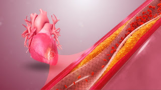
What causes coronary artery disease?A 3D illustration of a clogged artery
CAD is caused by a process known as atherosclerosis, in which plaque builds up in the inner walls of arteries, causing them to narrow and become rigid. This blocks blood flow, and the blockage can rapidly worsen if a piece of plaque breaks off and causes a blood clot.
One factor that increases the likelihood of atherosclerosis is the long-term presence of high concentrations of low-density lipoprotein (LDL) cholesterol in the blood. These high levels can result from a relative deficiency of LDL receptors, which are proteins on the membranes of various body cells, especially liver cells. LDL receptors capture LDL from the blood, causing its contents — cholesterol and fat molecules called triglycerides — to be taken inside the cell and out of the blood. Thus, a lack of these receptors causes LDL cholesterol levels to be higher in the blood.
High levels of LDL cholesterol provoke and exacerbate what doctors call atherosclerotic cardiovascular disease, an umbrella term for atherosclerotic changes that can develop in blood vessels throughout the body; this includes CAD, which specifically affects the coronary arteries.
Other factors that can provoke atherosclerosis include uncontrolled type 2 diabetes, hypertension (high blood pressure) and tobacco use.
Story continues
What are the symptoms of coronary artery disease?Man holding chest in pain.
Symptoms of coronary artery disease generally don't appear until the narrowing and hardening of coronary arteries begins to obstruct the flow of blood to the myocardium (the muscular layer of the heart).
When this obstruction does occur, it can produce angina — chest pain or chest heaviness that arises when the heart doesn't get enough oxygen — as well as pain in other places, such as the neck, shoulder or arm. Angina can be stable at first, meaning that it generally occurs only with exertion, related to physical activity or emotional stress and the strength of each heart contraction. However, this can progress to unstable angina, which is a type of acute coronary syndrome (ACS), a range of conditions associated with sudden, reduced blood flow to a part of the heart.
Known colloquially as a "heart attack," ACS is a spectrum running from its least severe subtype, unstable angina; to an intermediate subtype, non-ST elevation myocardial infarction (NSTEMI); to its most severe subtype, ST elevation myocardial infarction (STEMI).
According to the Mayo Clinic, symptoms of ACS resulting from coronary artery disease may include:
Dyspnea (difficulty breathing or shortness of breath)
Chest pain or chest heaviness
Pain in other places, such as the neck, shoulder or arm
Fatigue
Palpitations
Dizziness or fainting
Nausea
Sudden sweating
female doctor taking a man's blood pressure
To diagnose coronary artery disease, doctors will look at a patient's medical history and any symptoms that could indicate heart problems. They may also perform a physical examination.
Often, they will request an electrocardiography (ECG) test. ECG is a technique that detects changes in voltage over time across the heart, at different angles. When CAD causes enough blockage of blood flow to cause ischemia (insufficient blood supply) in particular parts of the heart, ECG can show abnormalities in the heart's activity.
Doctors also will request blood tests. Important blood values include the concentration of LDL cholesterol in a person's blood and the concentration of triglycerides (fat molecules).
Patients may be referred to a cardiologist for further tests, such as additional ECG testing, ultrasound imaging of the heart (echocardiography), or evaluation with a wearable heart monitor, a version of ECG that you wear for a few days or weeks that transmits data to the cardiologist.
Complications of coronary artery diseaseCoronary artery disease can lead to a condition called myocardial ischemia in which an artery cannot deliver adequate quantities of blood and oxygen to the myocardium. This can happen due to an artery gradually becoming blocked with plaque, resulting in the lumen of the artery (the hollow middle through which blood passes) narrowing and the wall of the artery hardening.
In addition, an atherosclerotic artery that has not been blocked enough to cause ischemia can suddenly become blocked by a piece of plaque breaking off, getting stuck and growing larger by stimulating the clotting process. Generally, this would lead to ACS.
Computer monitor showing cardiac stress test results of a senior male patient in ECG bike. Close-up of computer screen in cardiology clinic.
Even without a plaque rupture, however, the gradual increase in blockage driven by atherosclerosis disrupts a person's ability to perform physical activities, such as walking up stairs or around the block.
When doctors suspect that a patient's CAD may be causing life-threatening problems with the heart, such as ACS, myocarditis (inflammation of the heart's muscular tissue) or pericarditis (inflammation of the sac surrounding the heart), blood is tested to see if levels of certain enzymes are elevated, including a group of enzymes called troponins. Troponins are measured because they leak out of heart muscle tissue when it is damaged.
In cases of ACS, doctors can use troponin measurements and ECG to distinguish between broad subtypes of ACS, which has important implications for treatment and further testing. In cases when either heart failure or myocarditis is suspected, they'll also check blood for levels of brain natriuretic peptide (BNP), a hormone that enters the blood when the heart is stressed.
Treatment for coronary artery diseaseTreatment for CAD usually involves a combination of medication and lifestyle changes.
Medications can include cholesterol-lowering drugs, aspirin (to make it harder for platelets to stick to one another and to walls of blood vessels to form clots), medications to slow the heart while increasing the power of each contraction, or medications to widen blood vessels and/or to decrease blood pressure.
RELATED STORIES
—9 heart disease risk factors, according to experts
—Why are heart attacks more common in winter? A cardiologist explains
—Do other animals get heart attacks?
Lifestyle changes can also help to reduce a patient's risk of CAD and related health problems. According to the Cleveland Clinic, these include stopping smoking, limiting alcohol use, maintaining a healthy weight, limiting foods high in saturated fat, sodium and sugar, and getting enough sleep.
The Eye And Acute Coronary Syndrome: Getting To The Heart Of The Matter
Japanese investigators have found an ocular biomarker of cardiovascular diseases.
(Image Credit: AdobeStock/ArLawKa)
Japanese investigators, led by Taiji Nagaoka, MD, PhD, have found an ocular biomarker of cardiovascular diseases. Specifically, in a recent study, the mean blur rate (MBR) in the nasal region was associated with systemic atherosclerosis in patients with acute coronary syndrome (ACS).
Dr. Nagaoka reported the findings at the Association for Research in Vision and Ophthalmology annual meeting. He is associate professor, Department of Ophthalmology, Nihon University School of Medicine, Tokyo.
This team of investigators reported previously the retinal blood flow parameters were associated with systemic atherosclerosis in patients with ACS. What remained unclear was the relationship between atherosclerosis and the choroidal blood flow parameters.
In a continuation of their research, the team used laser speckle flowgraphy (LSFG) to study the association between the choroidal blood flow and the systemic risk factors for atherosclerosis, ie, the brachial-ankle pulse-wave velocity (baPWV), an index of systemic arterial stiffness, and the intima-media thickness (IMT) in patients with ACS.
The study evaluated 44 patients between April 2019 and September 2020. The patients underwent a lipid panel study, renal function tests, cardiac function tests, measures of the INT and baPWV, and an ophthalmic evaluation.
Dr. Nagaoka explained that they used LSFG to measure the choroidal MBR as an index of choroidal blood flow nasally (MBR-N) from the disc without large retinal vessels and compared the results with the systemic risk factors including the baPWV and IMT.
"The results showed significant positive correlations between the baPWV and choroidal blood flow nasally (r=0.33, p=0.029), between the LVDd and choroidal blood flow temporally(r=-0.30, p=0.045), and between the LVmass index and the retinal blood flow (r=-0.45, p=0.002).
The study reached the following conclusions. The choroidal MBR in the nasal region is correlated with systemic arterial stiffness and is significantly lower in patients with coronary artery multivessel disease. Evaluation of the ocular blood flow using the noninvasive LSFG technology can detect systemic risk factors in patients with ACS.
World-first Vascularization To Advance Global Research Into Heart Disease
Australian researchers have achieved two firsts that will assist in the global battle against heart disease: they created a tiny beating heart with its own vascular system and then uncovered how the vascular system affects inflammation-driven heart damage.
Cardiovascular diseases (CVDs) are one of the leading causes of death globally. According to the World Health Organization (WHO), CVDs claim an estimated 17.9 million lives yearly. Death rates due to CVDs are expected to rise, given our aging population and the impact of lifestyle-related risk factors.
CVDs include any condition that affects the heart or circulation, such as heart attack and coronary artery disease, high blood pressure, stroke, and vascular dementia. Given the prevalence of CVDs, it's important that research continues to uncover new ways of preventing, diagnosing and treating this group of diseases.
Australian researchers have contributed to the acceleration of research in the area of heart disease with their creation of a tiny heart organoid.
Organoids are tiny structures that mimic human organs. They're grown in a lab, using human pluripotent stem cells, which can be generated using 'reprogrammed' skin or blood cells.
"Each organoid is only about the size of a chia seed, measuring just 1.5 millimeters [0.06 in] across, but inside are 50,000 cells representing the different cell types that make up the heart," said James Hudson, corresponding author of the study.
Here, researchers created a tiny beating organoid, which is nothing new. But, for the first time, they were able to successfully incorporate vascular cells, the cells that line blood vessels, bringing the model heart even closer to replicating the real thing.
"Incorporating the vascular cells for the first time in our mini heart muscles is very significant because we found they had a key role in the biology of the tissues," Hudson said. "Vascular cells made the organoids function better and beat strongly. This has really opened up our ability to better understand the heart and accurately model disease."
The added bonus of vascular cells meant that the researchers could investigate how they affect inflammation, which can cause the heart to stiffen. In another first, the researchers uncovered the key role the vascular system plays in inflammation-driven heart muscle injury.
"When we stimulated inflammation in our mini heart muscles, we found the vascular cells played a central role," said Hudson. "We only saw the stiffening in the tissues that had the vascular cells. The cells sensed what was happening and changed their behavior, and we identified that the cells release a factor called endothelin that mediates the stiffening."
The researchers say that this discovery, and the use of their novel heart organoid, could lead to new treatments for heart disease.
"That's where our new system of producing vascularized cardiac organoids will really give us an advantage because we'll be able to progress the search for new treatments much more quickly," Hudson said.
Publication of the study will help researchers worldwide create their own vascularized organoid, boosting the global effort to tackle heart disease, the researchers say. Moreover, they say their discovery could be used to create kidney and brain organoids, accelerating research into the diseases that affect those organs.
The study was published in the journal Cell Reports, and the below video from the QIMR Berghofer shows the novel human heart organoid in action. James Hudson, one of its creators and authors of the current study, explains how the organoid was created and how it might be used.
Vascularised heart organoids video V2
Source: QIMR Berghofer


Comments
Post a Comment